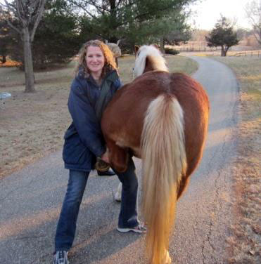
Lameness Examinations
Lameness in the horse is defined as an alteration in the horse’s gait or way of moving. This alteration could be from a variety causes. A few examples include laminitis, hoof abscess, strain or sprain of a tendon, ligament, or muscle, arthritis, fracture, OCD or chip within a joint, conformational or developmental related lameness, and a variety of other causes.
Typically a lameness exam begins by the veterinarian asking the owner questions about the horse’s history, such as when the lameness was first noticed, and what signs of lameness the horse has been exhibiting. The owner may have noticed an obvious head bob or shortening of stride, or simply a decrease in performance such as not wanting to take a lead or less bend going one direction. The veterinarian may palpate certain areas of the horse and place a hoof tester on the horse’s hooves. This is called the passive part of the lameness exam. Typically, there is some degree of an active exam where the veterinarian asks to see the horse move at a walk, trot, or canter in either a straight line or circle. The veterinarian may attempt to exasperate the lameness, or make it more apparent, by performing flexion tests of the limb joints. If an obvious lameness is present, the veterinarian may place a local nerve block or joint block. The purpose of an anesthetic block is to localize the source of the lameness. If the horse goes sound after a block is administered, the lameness can be localized to the area at or below the block.
Depending on the results of the lameness exam, your veterinarian may recommend further diagnostics such as radiographs or ultrasound. Treatments may also be recommended such as anti-inflammatories, joint supplements, joint injections, shockwave, or chiropractic care. Look for more information about these treatments under their respective links on our services page.

